
Curr Health Sci J, vol. 51, no. 4, 2025
AQP4 drives gliotic changes in an APPPS1 mouse model of Alzheimer’s Disease
[Original paper]
M.D. MANESCU(1), B. CATALIN(2), V.O. MATEESCU(1), G.C. ROSU(1), I.K.S. BOBOC(2), A. ISTRATE-OFITERU(1), I.M. LILIAC(1), C.J. BUSUIOC(1), D. PIRICI(1)
(1)Department of Histology, University of Medicine and Pharmacy of Craiova, Romania;
(2) Department of Physiology, University of Medicine and Pharmacy of Craiova, Romania
Abstract:
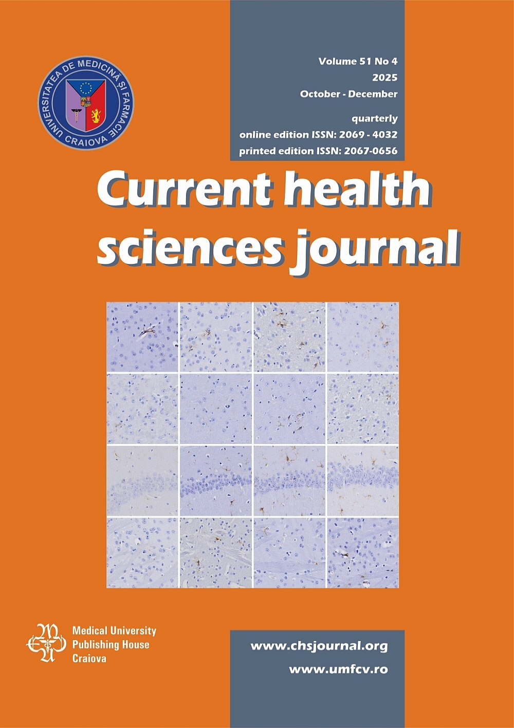
Alzheimer’s disease (AD) is the most common form of dementia, accounting for most of the cases, especially in individuals aged 65 and older. While the existence of genetic factors has helped us create animal models of AD that mimic APP and Ab overproduction, sporadic cases represent the bulk casuistry, and most probably their pathology is related to a loss of function towards the clearance of Ab rather than consecutive the overproduction of Ab alone. It is known that aquaporin 4 (AQP4) facilitation amplifies the perivascular Ab clearance route and decreases Ab deposits in animal models of AD, however it is now known how the glial component of the CNS responds to this treatment. We have aimed here to assess the glial response in a APPPS1 transgenic mouse model of AD, after one month of pharmacological facilitation or inhibition of AQP4. To this extent, we have utilized APPPS1 mice of 2 months of age, treated daily for 28 days with either TGN-020 AQP4 inhibitor or the TGN-073 AQP4 facilitator, and compared their GFAP expression in the brain with that of untreated APPPS1 and wild-type animals. Our image analysis of the GFAP immunohistochemical pattern, showed that AQP4 facilitation increases GFAP expression in the brains of APPPS1 animals, compared to untreated APPPS1 animals, but complexity-lacunarity morphological patterns resemble in fact those of wild-type animals, rather than that of the APPPS1 untreated animals, suggesting that this GFAP reactivity might represent a benefic amyloid-clearance astrocytic profile.
Keywords: Aquaporin 4, Alzheimer’s disease, mouse model, amyloid plaque, APPPS1
Corresponding: Valentin Octavian Mateescu, Department of Histology,University of Medicine and Pharmacy of Craiova, Romania, e-mail: valentin.mateescu@umfcv.ro; Pirici Daniel, Department of Histology, University of Medicine and Pharmacy of Craiova, Romania, e-mail: daniel.pirici@umfcv.ro
DOI 10.12865/CHSJ.51.04.13 - Download PDF AQP4 drives gliotic changes in an APPPS1 mouse model of Alzheimer’s Disease PDF
 Download cover
Download contents
Journal archive
- vol. 52 no. 1, 2026
- vol. 51 no. 4, 2025
- vol. 51 no. 3, 2025
- vol. 51 no. 2, 2025
- vol. 51 no. 1, 2025
- vol. 50 no. 4, 2024
- vol. 50 no. 3, 2024
- vol. 50 no. 2, 2024
- vol. 50 no. 1, 2024
- vol. 49 no. 4, 2023
- vol. 49 no. 3, 2023
- vol. 49 no. 2, 2023
- vol. 49 no. 1, 2023
- vol. 48 no. 4, 2022
- vol. 48 no. 3, 2022
- vol. 48 no. 2, 2022
- vol. 48 no. 1, 2022
- vol. 47 no. 4, 2021
- vol. 47 no. 3, 2021
- vol. 47 no. 2, 2021
- vol. 47 no. 1, 2021
- vol. 46 no. 4, 2020
- vol. 46 no. 3, 2020
- vol. 46 no. 2, 2020
- vol. 46 no. 1, 2020
- vol. 45 no. 4, 2019
- vol. 45 no. 3, 2019
- vol. 45 no. 2, 2019
- vol. 45 no. 1, 2019
- vol. 44 no. 4, 2018
- vol. 44 no. 3, 2018
- vol. 44 no. 2, 2018
- vol. 44 no. 1, 2018
- vol. 43 no. 4, 2017
- vol. 43 no. 3, 2017
- vol. 43 no. 2, 2017
- vol. 43 no. 1, 2017
- vol. 42 no. 4, 2016
- vol. 42 no. 3, 2016
- vol. 42 no. 2, 2016
- vol. 42 no. 1, 2016
- vol. 41 no. 4, 2015
- vol. 41 no. 3, 2015
- vol. 41 no. 2, 2015
- vol. 41 no. 1, 2015
- vol. 40 no. 4, 2014
- vol. 40 no. 3, 2014
- vol. 40 no. 2, 2014
- vol. 40 no. 1, 2014
- vol. 39 no. 4, 2013
- vol. 39 no. 3, 2013
- vol. 39 no. 2, 2013
- vol. 39 no. 1, 2013
- vol. 38 no. 4, 2012
- vol. 38 no. 3, 2012
- vol. 38 no. 2, 2012
- vol. 38 no. 1, 2012
- vol. 37 no. 4, 2011
- vol. 37 no. 3, 2011
- vol. 37 no. 2, 2011
- vol. 37 no. 1, 2011
- vol. 36 no. 4, 2010
- vol. 36 no. 3, 2010
- vol. 36 no. 2, 2010
- vol. 36 no. 1, 2010
- vol. 35 no. 4, 2009
- vol. 35 no. 3, 2009
- vol. 35 no. 2, 2009
- vol. 35 no. 1, 2009